Centre for Bioimaging

 News

Check out our new publication in Nature Comminications, focused on volume electron microscopy of the plasmodium microgametocyte. Lots of beautiful SBF-SEM and serial EM tomography data and models of a key stage in malaria transmission.

About us

The Oxford Brookes Centre for Bioimaging is a core facility based in the Faculty of Health and Life Sciences.

Our unit has a wide range of light and electron microscopes, as well as fully-equipped electron microscopy sample preparation labs.

We also have dedicated workstations for 3D image analysis. Our expertise lies in high-resolution live cell imaging and 3D electron microscopy, also known as volumeEM (vEM).

What we do at the Centre for Bioimaging

Light Microscopy:

  • Widefield imaging of live and fixed samples
  • Confocal microscopy
  • Fluorescence live cell imaging
  • Super resolution confocal microscopy (Airyscan)
  • Whole organism fluorescence microscopy
  • Expansion microscopy

Electron Microscopy:

  • Scanning electron microscopy (conventional SEM)
  • High-resolution SEM 
  • 3D SEM (serial block-face SEM)
  • Thin-section transmission electron microscopy (TEM)
  • Serial section TEM (ssTEM)
  • TEM serial tomography of plastic sections
  • Immunolabelling for TEM and high-resolution SEM
  • Conventional (chemical fix) sample preparation for EM 
  • Cryo fixation for EM (high pressure freezing and freeze substitution)
  • Correlative light and electron microscopy (CLEM)

Research impact

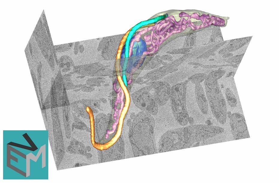
Volume Electron Microscopy 

We are experts in 3D electron microscopy, also known as volume EM (or vEM). 

Volume EM has had a huge impact in all areas of biological and medical research, by adding a new dimension to our understanding of the biology of cells and tissues.

We perform vEM techniques routinely, including serial block-face scanning electron microscopy (SBF-SEM) and serial electron microscopy tomography. Our expertise in vEM is the source of numerous collaborations between our researchers and other institutions around the world.

We are part of the vEM commmunity initiative and are working together with other vEM centres to exchange knowledge and improve techniques. As part of this effort, we organise the SBF-SEM focus interest group, to help the community share tips and tricks and keep up with developments in the field of SBF-SEM.

volume EM

Access and booking

Access

The Centre for Bioimaging is open to University staff and students, as well as to external users. We provide services and/or training, as required. We can provide assistance in designing microscopy experiments. All projects require safety evaluation/approval by the Bioimaging team.

To access the centre, please contact bmsbioimaging@brookes.ac.uk.

Booking

If you already have access to Oxford Brookes Centre for Bioimaging, you can use our booking system to book equipment. You will need your Calpendo login and password.

If you do not have access via Calpendo, or have a general enquiry, please contact bmsbioimaging@brookes.ac.uk.


Book equipment

Meet the team

Professor Sue Vaughan

Professor Sue Vaughan

Professor of Cell and Molecular Biology

Dr Flávia Moreira-Leite

Dr Flávia Moreira-Leite

Bioimaging Manager / Electron Microscopy Specialist

Dr Alan Marron

Dr Alan Marron

Bioimaging Technician / Light Microscopy specialist